
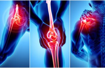
Computed tomography (CT), now and then referred to as “computerized tomography” or “computed axial tomography” (CAT), is a noninvasive medical exam or system that uses a specialized X-ray device to supply go-sectional pix of the body. Each move-sectional picture represents a “slice” of the person being imaged, like the slices in a loaf of bread. The best diagnostic center uses cross-sectional pictures for the diffusion of diagnostic and healing functions.
CT scans may be performed on every location of the body for a spread of reasons (e.g., diagnostic, treatment making plans, interventional, or screening). Maximum CT scans are completed as outpatient strategies.
How does a CT machine work?
• A motorized desk actions the patient thru a round opening within the CT imaging machine.
• At the same time as the affected person is inside the beginning, an X-ray supply and a detector assembly inside the device rotate around the affected person. A single rotation generally takes a 2d or less. During rotation, the X-ray source produces a narrow, fan-shaped beam of X-rays that passes through a phase of the patient’s body.
• Detectors in rows opposite the X-ray supply sign up the X-rays that bypass the affected person’s body as a snapshot inside the system of making a picture. Many exclusive “snapshots” (at many angles through the patient) are accumulated during one complete rotation.
• For every rotation of the X-ray source and detector meeting, the photo statistics are despatched to a computer to reconstruct all of the character “snapshots” into one or multiple move-sectional photographs (slices) of the inner organs and tissues.
CT pics of internal organs, bones, soft tissue, and blood vessels offer more readability and more information than traditional X-ray pics, including a chest X-Ray. You can check online for the CT scan center near me for the scanning.
An ultrasound is a safe, usually non-invasive medical imaging and diagnostic device. Other names for ultrasound include sonogram and ultrasonography at the ultrasound center in Noida.
Ultrasounds use sound waves to generate pix of the inside of your frame, allowing radiologists and your health practitioner to look at your organs, tissue, and other structures.
They do not use radiation like X-rays and different medical imaging technology. Widely, ultrasounds are used for one motive:
• Pregnancy ultrasounds study an unborn toddler and test its health and boom repute.
• Diagnostic ultrasounds offer statistics about internal regions and elements of the frame, which include the liver, kidneys health, and reproductive organs.
When they are an effective and precious tool within the right place and under the steerage of experienced, certified professionals, there are shortcomings.
How does ultrasound scanning work?
Ultrasound uses sound waves to supply black-and-white pictures of the body’s internal structures. Waves are produced by using a wand-like tool known as a transducer, which usually makes use of ceramic crystal materials called piezoelectrics.
Piezoelectrics create sound waves while an electric subject is applied and create an electric powered subject while sound waves are applied.
This allows the transducer to detect waves that leap off barriers between systems in your frame — for an instance, the boundary between tissue and bone.
The scanner measures the velocity and timing of each pondered sound wave. It uses this data to determine the space between the transducer and the boundary. Those distances are what generate pix. You can search in google for a digital X-ray near me for the digital x-ray.


